AI uses chest X-rays to identify fatty liver disease
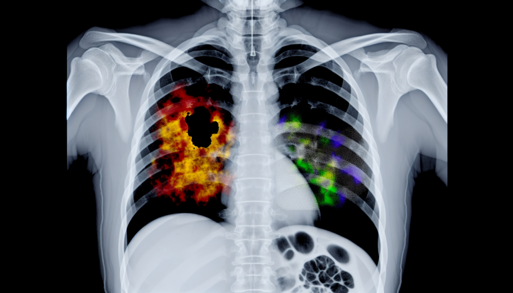
Image:
Chest radiographs, typically used to examine the lungs and heart, also display portions of the liver, enabling AI to identify indications of fatty liver disease.
Image credit: Osaka Metropolitan University
Fatty liver disease, a condition where fat builds up in the liver, affects roughly 25% of the global population. Without treatment, it can progress into serious health problems such as liver scarring or cancer, highlighting the importance of early diagnosis and medical intervention.
Traditional methods for diagnosing fatty liver disease involve tools like ultrasound, CT scans, and MRI, which are often expensive and require specialized medical equipment. By contrast, chest X-rays are more commonly available, cost-effective, and involve minimal radiation. While their primary function is to evaluate the lungs and heart, they inadvertently capture part of the liver, presenting an untapped opportunity for disease detection. Despite this, the potential of chest X-rays in identifying fatty liver has not been widely examined.
To address this gap, researchers led by Associate Professors Sawako Uchida-Kobayashi and Daiju Ueda at the Graduate School of Medicine at Osaka Metropolitan University have created an artificial intelligence model capable of recognizing signs of fatty liver disease from chest X-ray images.
In this study, researchers analyzed 6,599 chest X-ray images from 4,414 patients, developing their AI model using data guided by controlled attenuation parameter (CAP) scores. The model demonstrated strong diagnostic performance, achieving an area under the receiver operating characteristic curve (AUC) between 0.82 and 0.83.
“Using standard chest X-rays, which are easy to access and low in cost, offers a promising new approach to diagnosing fatty liver disease,” said Professor Uchida-Kobayashi. “We hope this technique will eventually be adopted in routine healthcare settings.”
###
About Osaka Metropolitan University
One of Japan’s largest public institutions, Osaka Metropolitan University is dedicated to advancing society by combining diverse fields of knowledge and spearheading pioneering research on a global scale.
Journal
Radiology Cardiothoracic Imaging
DOI
10.1148/ryct.240402
Method of Research
Imaging analysis
Subject of Research
People
Article Title
Performance of a Chest Radiograph-based Deep Learning Model for Detecting Hepatis Steatosis
Article Publication Date
20-Jun-2025