Graz University of Technology pioneers fresh pathways in lung cancer study through digital cell twin innovation
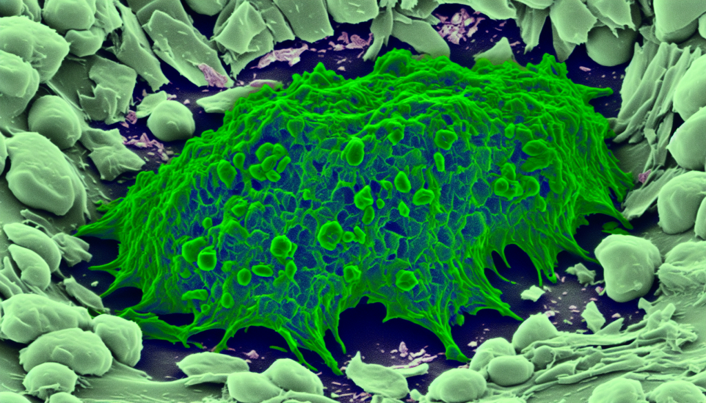
Image:
Scanning electron microscope image of a lung cancer cell.
Image credit: Anne Weston, Francis Crick Institute
A research team led by Christian Baumgartner from the Institute of Health Care Engineering at Graz University of Technology (TU Graz) has created an advanced digital replica of the A549 lung cancer cell line. This digital twin focuses on detailed simulations of internal bioelectric functions and calcium signaling processes. Calcium plays a critical role in cellular function and survival; however, high concentrations within a cell can trigger cell death. This property makes calcium a compelling subject for cancer research. Developed through the DigLungCancer project, this model elaborates on a previous 2021 version, the first digital ion current model for human A549 lung adenocarcinoma cells. The updated model opens new research paths into how calcium signaling and electrical charges across the cell membrane influence cancer cell development. Although these cancer cells do not respond to stimulation in the same way as nerve cells, they do exhibit measurable electrical behavior. The model offers the most comprehensive digital representation of cancer cell bioelectric processes so far. With long-term goals including the discovery of new drug targets and the development of computer-driven personalized therapies, the project receives support from the Styrian division of Austria's cancer support group, Österreichische Krebshilfe.
Advancements in Cell Cycle Analysis
“One of the major improvements in our new model is the refined simulation of how calcium is distributed inside cancer cells,” says Christian Baumgartner. “For the first time, we included localized areas—microdomains—where calcium levels are elevated, and where internal cell frameworks closely approach the cell membrane. In these zones, CRAC channels—calcium release-activated calcium channels—control calcium entry, which is vital for activating the signaling mechanisms that govern the cell cycle.”
This breakthrough allowed for an unparalleled depiction of electrical characteristics in cancer cells, capturing subtle activities such as calcium storage, transport systems, buffering ability, and the localized spread of calcium. At its foundation, the model integrates hundreds of mathematical formulas to generate simulations. With this digital model, researchers can examine how various pharmaceuticals affect cellular functions. For example, the model makes it possible to forecast how changes in calcium flow or ion channel behavior influence cancer cell proliferation or death. When predictions from the simulations are promising, they can undergo lab testing. Additionally, it allows for exploration of combinations of ion channel modifications, which would be nearly impossible or too labor-intensive to test experimentally. Already, the team has proven that blocking specific CRAC channels can reshape calcium distribution patterns within the cell and impact related signaling pathways—potentially leading to halted cell division or prompting programmed cell death.
Future Plans: Modeling Intercellular Interactions
Currently, the model represents only a single lung cancer cell. As a result, it does not yet simulate how cells interact with each other, form tumors, initiate metastatic spread, or promote new blood vessel formation in tumors. These topics are part of future phases of the research. Over time, this approach could make it possible to tailor simulations to different cell types or individual patient data, supporting personalized cancer diagnostics and therapy. The team believes this computational approach could extend beyond lung cancer to study other types, such as breast and prostate cancer.
Journal
Clinical and Translational Medicine
DOI
10.1002/ctm2.70456
Method of Research
Computational simulation/modeling
Subject of Research
Cells
Article Title
Computational modeling and simulation in oncology
Article Publication Date
5-Sep-2025